Featured

KEEP THAT GAMEFACE ON AND FIGHT LIKE A PRO

We are all confused souls waiting to find the reason for our lives,willing to use all our chances before we get consumed by the black hole.I was once “your answer to the matrix”,(my personal description on social sites.)now I am stuck in the matrix myself.

But we evolve,and that’s how we survive.We don’t stop and that’s how we grow.When we are given colors we scribble into our imaginary world; when we hold pens we make the best bachelors ,when we start working we accomplish task and when we get to share our experience we have stories to tell.

This post stems from a problem for which we have to find a solution. Looking at the facts,we found tooth loss in people under 35 is most commonly caused by athletic trauma,fights or accidents.A non expensive tool that is available and is a possible way to protect your face from many injuries that your teeth and jaw suffers while playing, gym training on the ground and practicing contact sports. The injuries that require life long care and expenses and could be easily averted had we been aware and asked questions.

The National youth sports foundation for the prevention of Athletic injuries has
reported that dental injuries are the most common type sustained during participation in sports.Most of these injuries can be reduced or at least prevented with the use of proper equipment like a guard.A mouth guard is a soft plastic or laminate device used in sports to prevent oral injuries to the teeth,mouth, cheeks, tongue and jaw.According to different reports, sports-related facial fractures account for 20% of all sports injuries and as much as 33% of all facial bone fractures.The use of a mouth guard can prevent more than 200,000 oral injuries to the mouth each year.

This post intend to create awareness towards parents and the youth that protecting your teeth and jaws are your absolute right and every school should make mouth guards a mandatory requirement for all contact sports and training.It is as much a school responsibility as of the parents when their children opt for contact sports as activities. All contact sports like basketball, baseball, wrestling,mixed martial arts,soccer and volleyball may consider wearing a mouth guard to prevent injuries to the mouth.Just like camps for dental checkup and medical vaccination,schools can coordinate with their local dentist for mouth guard impression every new semester and bring them into continual care and safe sports.

For me it will always be “no guard no play”.All that is required is you fixing up an appointment with your dentist specialized in making mouth guards for your requirement. Over the counter available mouth guards are “one size fits all” can not provide adequate protection because your jaw is not like others.

“You can prevent accidents or you can pay for a broken smile”

Can A Mosquito Cause Bleeding Gums?

Dengue fever (DF) is the condition of toxic hemorrhagic viral disease transmitted by a female Ades mosquito. The incubation period of the virus after the mosquito bite is between 3-8 days. The clinical features vary according to the age of the patient and generally start as a flu-like illness .Infants and young children usually have mild to moderate symptoms like a high fever with a rash that is hard to distinguish from other viral illnesses. If left undiagnosed it may take a more severe form like Dengue Shock Syndrome or Dengue Hemorrhagic Fever having an increased mortality. Other symptoms may include pain of muscle, joint and behind the eyes along with nausea and vomiting. Currently India reported at least 50 deaths within a week in the month of August itself; These included mostly children in the northern part of the country. Going by the reports, children were more susceptible due to exposure in the playground and almost five times more likely to die during a second infection.


Bleeding contributes to worsening morbidity by the acute toxic state that appears during the 3rd to 5th day of illness following the onset of fever and usually last for 7 days. The most common hemorrhagic manifestation are bleeding nose, skin hemorrhages, scattered petechiae, ecchymoses and gastrointestinal hemorrhages and that included spontaneous bleeding from the gums and a purpuric rash. Most of these cases reported with declining platelet count, a condition knows as for thrombocytopenic purpura with the numbers as low as 2,000. (normal range :150,000 to 400,000).Laboratory tests are positive for immunoglobulin M (IgM) antibodies to the virus. Treatment includes managing fever and dehydration to administration of steroids for up to 10 days and platelet transfusion in the most sever form.


Areas of the world where dengue fever is common, one vaccine (Dengvaxia) is approved for people ages 9 to 45 who have already had dengue fever at least once. The World Health Organization however stresses that the vaccine alone is not an effective tool .In people who have not had dengue fever in the past (seronegative), receiving the vaccine appears to further increase the risk of severe dengue fever.


The disorders of blood or blood forming organs can have a profound effect on gums and bones of the jaw. Ecchymosis and petechiae observed most often in the soft palate area is a sign of an underlying bleeding disorder. Abnormal bleeding from the gums or other areas of the oral mucosa that is difficult to control is an important clinical sign suggesting a hematological disorder. The prompt referral of the patient may save the life of patient and thereby also emphasizes that the dentist can be the first person who can actually diagnose and refer these patients for proper management.

Self-driven Jaw Expanders in Special Needs

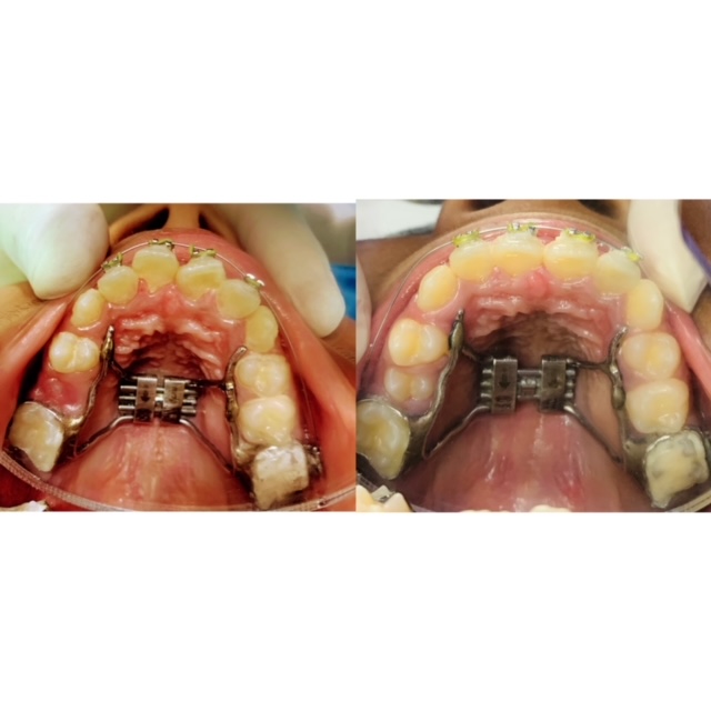
Rapid Maxilla Expansion

Your upper jaw bone forms in two halves joined together by a palate suture. These sutures close as early as when a patient reaches 12–13 years of age. Once the adult bone structure is set it can no longer undergo expansion unless it is assisted by a surgeon. The chances of successfully expanding the maxillary bony base, therefore, decline with the patient’s age. When the arch is narrow the loss of space leads to crowding of teeth with the eruption of the lateral incisors and canines into irregular alignment. However, if we could widen the upper jaw bone it will lead to more space dentition. As a result of which problem of crossbite, crowding, malalignment, abnormal positioning of teeth could be averted well in time.

The cause of malalignment such as crossbite can be due to associated habits such as nonnutritive sucking, airway obstruction leading to mouth breathing, lowered tongue posture, and short duration of breastfeeding. It is also commonly seen among children of Down syndrome and cerebral palsy given to their hypotonia (decreased muscle tone) of the tongue. Prevalence of crossbite ranges from 6 to 30% in the general population, but since it is not a self-correcting condition it should be treated as soon as it is diagnosed by means of maxillary expansion devices. The ideal age for a child to get a palate expander is when they’re young, around 7 to 8 years old. The palate is forming rapidly, so it’s easier to reshape.

RME Rapid Maxillary Expansion (sometimes called rapid palatal expansion) is a technique used to widen (expand) your upper jaw (palate) when it is narrow. The expander appliance is a fixed brace made from a mould of your teeth to fit your mouth only. It is fixed to the upper teeth. You cannot remove it without your dentist’s help. The RME moves these two halves apart and holds them there while new bone fills in the “gap” under the gum. The metal framework contains an expansion screw in the middle to widen the upper jaw. It is attached to metal bands cemented on the back teeth or to a plastic splint which is cemented firmly to the back teeth. The expansion screw in the metal framework needs to be turned, usually twice a day or as instructed by your dentist for which a special key is given to you for this purpose.

In young patients with special needs and those who must periodically undergo MRI ( such as epilepsy, autism, or vascular problems) or for whom it would be difficult to self activate a palatal expander due to muscle weakness or compliance problems and even those with allergy or hypersensitivity to metal; a metal-free fixed maxillary expander will be advantageous. The new concept of “self-driving orthodontics” is the new goal of future orthodontics which utilizes a fully pre-programmed digital analysis along with project printing and includes digitally monitored changes during and after therapy. The design of the device starts from an intraoral scan. It requires an initial virtual setup of the STL files for expansion of the upper arch; after the project is generated the relative STL file is used for the milling process (for PEEK) and 3D printing (for PA12) of the product appliance.  

The RME appliance has to be worn for a period of 3-9 months. In the initial course of 2-3 weeks, the upper jaw bone widens (up to 6mm). In the remaining months, it is left alone to allow new bone to fill in the gap under the gum between the two halves of the upper jaw. Treatment is mostly completed with a fixed brace to straighten your teeth. The Expander, due to its shape memory, progressively expands like a compressed spring in a very controlled and comfortable way.

The new device holds a promise to rectify jaw discrepancy and arch alignment among children with special needs without much intervention and need not be removed in case of radiographic procedure or MRI.

Good Gut Bugs Good To Replace Oral Bugs

Eric Metchnikoff proposed that people who consumed fermented food containing lactic acid bacteria lived longer and stronger. No guesses on why people from Bulgaria, Korea have a healthy and longer life. Good bacteria can eliminate toxins and free radicals that can damage skin and cause early signs of aging. They can fight bad bacteria linked to leaky gut, inflammation throughout body, including blotchy and old-looking skin.


We are looking at the live bacteria when administered in an adequate amount in food can confer loads of health benefits to the host. These are called ‘Probiotics’ unlike antibiotics (one which kills bacteria)and can strengthen the immune system to combat allergies, stress, exposure to toxic substances, and other diseases. In the oral cavity, they can alter the surrounding environment by first integrating into the salivary biofilm, then modifying the pH, and finally compromise the ability of other harmful pathogens to get established. Indirectly they can enhance local immunity and cause alteration in mucosal permeability acting as antioxidants, modulate the inflammatory response by producing antimicrobials like lactic acid, hydrogen peroxide, and bacteriocin. They will overall decrease the colony counts of harmful pathogens in the oral cavity by competitive inhibition.


The most commonly used probiotic can be yeast, bacteria, or molds. These include,
1)Lactic acid-producing bacterial (LAB) species like Lactobacillus, Bifidobacterium, Streptococcus.
2) Non-lactic acid Bacteria species like Bacillus, Propionibacterium
3)Nonpathogenic yeast like Saccharomyces

Kimchi from Korea contains pickled vegetables with loads of lactic acid bacteria, kefir contains yeast and our household yogurt contains billions of live cultures that can balance the ecosystem of gastrointestinal, genitourinary as well as oral cavity.

Our mouth is a niche to thousand of different species of bacteria. Of these elevated levels, Streptococcus mutans had long been associated with an increased risk of caries. Interestingly it was noted that regular consumption of yogurt, milk, or cheeses contains a probiotic which led to a decrease in the number of cariogenic streptococci in the saliva as well as a dental plaque for up to 80 % in 2 weeks. Also, probiotics containing L. rhamnosus and L. casei or Bifidobacterium have shown to decrease the growth of streptococcus. Cheese contains L.rhamnosus that can decrease the fungal outgrowth of Candida. Cheese has been reported to prevent demineralization of enamel and in turn, promotes remineralization. Researchers proved that 14 days intake of L.returi led to the establishment of the strain and also caused a significant decrease in diseases causing pathogens of gingiva and periodontium. These harmless bacteria could successfully reduce gum inflammation and plaque accumulation and positively showed improvement in gum health by lowering probing depth and even loss of attachment.


Probiotics were however delivered as food supplements and not drugs for a very long time. As a result of which direct action was not possible locally until the use of chewing gum, lozenges, tablets, and mouthwash gain acceptance. Mouthwash with L. reuteri , tablets of L.salivarius with Xylitol gum has shown to reduces plaque pathogens and promote the healthy ecological balance of useful bacteria like S.oralis and S. uberis. Mouth wash containing Weissella cibaria has shown to decrease fetid odor by removing methanethiol and hydrogen sulfide. Gum and lozenges containing S.salivarius have shown to decrease volatile sulfur components causing bad breath.


In India probiotic is mostly given in pediatric patients and some adults as Yakult, Sporolac, or Vibactum (VitaminB+ Bacillus) or with some antibiotic preparation. Only mild side effects have been noted to date with most of them associated with bloating and gas. Probiotics has been used sparingly in immunocompromised patients due to the associated risk of pathogenic infection. Clinical studies conducted so far suggest that probiotics could be useful in preventing and treating oral infections, including dental caries, periodontal disease, and halitosis. Therefore, further research is warranted in nonpathogenic bacterial strains with the aim to reduce cariogenic potential, longer duration of local action, and replacement to a healthy ecosystem for caries-free oral condition.

ABOUT THE CHILD WHO BLEEDS

Blood is a living tissue composed of solid and liquid parts. The solid part is made of white and red blood cells, and platelets. Disorder of blood can affect one or more of these parts and may lead to prolong bleeding episodes that are fatal in children if undiagnosed.

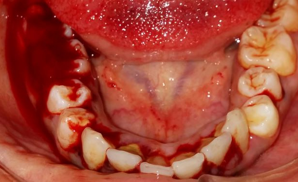
Consensus shows 80% Indian suffer from life threatening blood disorder that remain undiagnosed due to unavailability of diagnostic facilities. Bleeding from the mouth is the primary finding at age of 6to 12 months when the first tooth erupts or at the loss of first tooth. This raises the first red flag in the scenario of prolonged and voluminous bleeds in the oral cavity. Most common site of spontaneous bleeding will be gums, frenum of lips and tongue and will occur at the slightest provocation in the form of brushing, food abrasion or even infection. Oral cavity is highly vascular and therefore profuse hemorrhagic episodes are seen among children under emotional stress and extreme physical activities.

Epistaxis (bleeding from nose) is the common condition in preschool children and prolong episodes of bleeding follow even on the simple allergic rhinitis. While children with sharp teeth may bite on cheeks, oral ulcers and bruising is commonly reported in their lips and tongue. Management of dental trauma needs an immediate attention to reduce blood loss. Oral treatment should therefore be provided by an equipped staff trained to diagnose and control bleeding in an event of emergency.

Hemophilia and Idiopathic Thrombocytopenic purpura (ITP) are commonly prevalent bleeding disorders among children. Hemophilia is an inherited disorder where the blood fails to clot normally and may be attributed due to absence of any of 12 factors responsible for clotting but majorly factor8,9 & 11. Such children will bleed longer through nose, oral cavity, urine, muscles and joints. ITP on the other hand, is an autoimmune (self-destructing) disorder with unknown cause, characterized by decreasing platelet number. Platelet are cell responsible to form a clot and as a result of deficiency easy bruising, bleeding from the gums and internal bleeding are evident. It may have a sudden onset following a viral fever and usually disappear in few weeks, commonly seen among 2-6 years old.

Currently no universally acceptable standard protocol is available for affected children of blood disorder to ensure a safe treatment in clinical setting therefore both local and systemic management is advisable in collaboration with a hematologist (blood specialist).Children with Hemophilia will need a prerequisite of antifibrinolytics (clot thickener)and stabilizing agents(clot protectors) and even blood transfusion in an event of excessive blood loss .ITP will require steroids, and platelets transfusion in case of fatal internal bleeding due to trauma.

Dental management prefers roots canal treatment over extraction and splinting over removal of loose fragment. Periodontal therapy for the gum diseases must be initiated on a regular basis to avoid emergency situation later. Interestingly, poor oral care, diet and fear of bleeding are the most common reason seen for failure to observe oral health. Therefore, reducing dental fear and anxiety is a key to continued dental care.

Most children can have a normal independent life. Preventive measure in form of protective clothing, padded crib, protective shield/helmet, bite guard during active sports are beneficial. Contact sports may be practiced with caution and drugs that may interfere with the body’s ability to control bleeding must be avoided. Most oral and dental diseases can be prevented but dental treatment with children of blood disorder is complex and requires dedicated training.

SILVER LINING FOR SPECIAL NEEDS

Children exhibit high caries prevalence under the age of 2-7 years old. Such high caries activity is caused by the acid producing bacteria in the plaque like Scardovia wiggesie, Lactobacillus salivarius, Streptococus mutans and Parascardovia denticolens.To counter this bacterial ingress silver was found to be indispensable for very many years. Perhaps the earliest use of silver was in the form of dental amalgam.

Silver has shown chemical affinity and by virtue of its active ions they interact with the thiol group of protein and phospholipids parts of bacterial membrane. Silver ions adhere to cell wall and via electrostatic attraction they can disrupt the bacterial envelope. Once inside the cell the silver ions deactivate the respiratory enzymes, generate reactive oxygen species and inhibit protein synthesis. This causes problem in DNA replication and cell production, thereby structural changes and cell death.

The antimicrobial efficacy of silver ions is inversely related to particles size, that is, lesser the size means higher efficiency with higher bactericidal effects. Biomaterial technology has evolved second generation of silver ions as nanosilver and nanoscale silver nanoparticles. At such size and with greater surface area, silver interact more intensely with other oxygen species and inorganic molecules creating oxidative stress.

 The combine antibacterial effects of silver and the remineralizing effects of fluoride was seen in SDF , which is a promising therapeutic agent for managing caries lesions in young children and those with special care needs. The use of SDF is recommend for seniors who presented with increased risk of root caries. Silver in SDF (silver diamine fluoride) act as a potent germicidal and releases fluoride to reduce dental decay. Of the many concentration 38% SDF was found to be most effective and used commonly to prevent collagen degeneration and inhibit demineralization.38% SDF releases 44,800 ppm fluoride and was best known to arrest caries attack by increasing the micro hardness of dentine in primary teeth with the ability to form sclerotic dentine.

The major drawback of SDF is staining of carious tissues to dark black due to oxidation process of ionic silver. To overcome this drawback nano silver flourides(NSF) were developed. It is a colloid base on chitosan, silver nano particles and fluoride which prevent dental decay in children by inhibiting plaque and biofilm formation. Addition of silver nano into dental material like composite resin has shown to reduce bacterial count of S.mutans and L.bacillus. As a new anticaries agent silver nano was found to assert potent antimicrobial effect about 25 fold higher than chlorhexidine. As a dentifrice/toothpastes, 5% Nano-silver incorporated Sodium fluoride (NSSF) was 8 times more economical compared to SDF and hence its use at community level can be cost effective and reliable.

Silver nano particle have shown to reduce incidence of dental decay in milk teeth which are most sensitive to fluoride treatment because the enamel is 150 times more permeable than permanent teeth. The silver will tend to remineralize dentine and enamel by increasing fluoride uptake when acidic breakdown of enamel is increased by bacteria.

To meet the ever increasing demand of good oral health among underprivileged and special needs where meals are highly cariogenic  and oral care is difficult silver nano particles was found to be safe and equitable caries control agent that can be used to help meet WHO millennium goals and fulfill US criteria for 21st century medical care.

Efforts with the health sector should be made at global scale to promote highest level of evidence based dentistry with long term follow up for  making  conclusive recommendation on silver products. This could be a major breakthrough in preventive and therapeutic dentistry for those who need oral care and are underprivileged or require special care.

KID’S DENTIST FIGHT CANCER OF KIDS

Children with malignancy are at the increase risk of short and long term effects of radiation and chemotherapy. Treatment for cancer is directed mainly to rapidly dividing cells but children at the age of 2-3 years old have higher metabolic activity towards all the cells. This would mean the growth of actively dividing structures like bones, teeth and soft tissue are going to be hugely impacted.

Only radiation as low as 200cGy is microscopically evident to cause cellular damage, when children are subjected to 1800 cGy we are looking at irreparable damages with greatest cranial disturbances and anomalies amounting to lack of mid facial growth, loss of anteroposterior dimension and discrepancy of transracial height and orbital dimension. Chemotherapy adversely affects the metabolic hormone production leading to osteoporosis, thinning of bones and loss of bone density as a result of which increased susceptibility to fractures are noticed.

Most of the children diagnosed with cancer are at the age of 1-6 years. Tooth development begins in the 6 weeks of intrautrine life and continues till 14-15 years. Each tooth bud undergoes quantitative and qualitative differentiation during odontogenesis and any interferences during the cycle leads to defects in shape, structure, number and tooth eruption. When chemotherapy/ radiation coincides with dental development odontoblast necrotizes and their activity decreases, secretory function is impacted, membrane permeability diminishes and mineralization of tooth is affected causing loss of teeth and hypo mineralized structures. These changes are well observed as hypoplastic enamel patch.


Alteration in cell cycle extends to root development as the cytostatic drugs prevent the formation of Herwig Epithelial Root system( precursor for roots). The root aberrations are manifested as short , fragile roots and taurodonts (large crowns at the expense of roots).Root edification of deciduous(milk) teeth is evident with intensified resorption before their physiological age resulting in premature tooth loss effecting the sequence of eruption in permanent teeth. The severity of these damages will depend on the type of medication, dosage and frequency of therapeutic cycles, the age of the child at the moment of initiation of cancer therapy, and the stage of the dental development. Among the commonly used cytostatic agent is colchicine, vinblastine, cyclophosphamide, and methotrexate.


Damages of the soft tissues following immediate radiation (4000 cGy/rads or higher ) often occurs on the salivary glands due to alteration in their vascular supply. As a result of which xerostomia (dry mouth) ensues and the pH falls from 7.0 to 5.0, which is cariogenic, the buffering capacity of saliva decreases, consequently, remineralization capacity of saliva is hampered, and the minerals of enamel and dentin therefore dissolve easily.

With radiation as low as 40 Gy persistent sore throat, hoarseness, burning sensation in the mouth and gums, problems speaking, difficulty in swallowing, trismus( hardening of jaw muscles) make it difficult to open mouth due to scaring. The harm on the chemical and microbiological barrier function is devastating. Destruction of epithelial barrier may lead to extreme painful condition like oral mucositis and ulceration reducing nutritional intake in a stressed child.


Education of parents regarding short and long term effects in the oral cavity and craniofacial complex will provide emphasis on the importance of oral care. Radiation and cytostatic drugs both attack the periodontal status aggravating bleeding due to lower platelet count and damaged neutrophil functionality. Therefore, all local factors causing infection should be eliminated before therapy. Cancer therapy targets the bone marrow, suppressing the defense mechanism and causes reduced antibody and proteins to fight infections. Any existing lesion may flare up presenting a life threatening scenario when the child immunity is already compromised.


Quality of life of leukemic child is adversely affected and requires early intervention and definitive management. To reduce the risk of oral and systemic complication comprehensive oral hygiene measure should be established that could provide dental and oral care before initiation of cancer therapy, during immunosuppression and to manage post effect of the treatment. Teeth with poor prognosis should be removed ideally 3 weeks before cancer therapy to allow adequate healing and prevent the a risk of osteoradionecrosis. Endodontic management is severely compromised due to short roots. Orthodontics is restricted to removable appliances and retainers in order to minimize the risk of root resorption and to use lighter forces as the supporting bone are not strong enough for braces. Bonding of teeth and restorative treatment becomes a necessity for hypoplastic teeth prone to radiation caries. Oral pediatric medications are high on the sugar content which makes hypoplastic teeth susceptible to dental caries. A regular follow up and fluoride application will help to maintain the oral health for already compromised teeth.

The present health network is not supportive in providing adequate dental health and effective oral care. Here it is for the the benefit of community at large that a cooperation must exists between pediatric and dental clinic for easy referral and primary care. To avoid complications and to reduce morbidity following cancer therapy the presence of pediatric dentist in the oncology team must be made mandatory. Finally, health governing body must take significant initiative to improve oral care of children with leukemia.

Disability, Specially Needs Nourishment

One billion people worldwide are malnourished, and around one billion live with a disability. According to UNICEF 2006, 5-6 million children under age of 5 years contribute to mortality rate because of high prevalence of malnutrition. This accounts to 10 children/ minute. Most of these disabilities weather physical or sensory like deafness, blindness, intellectual and mental health are results from the nutritional deficiency at numerous points throughout life cycle.

Therefore, disability and malnutrition go hand in hand in description of cause and contribution per se. While the Right to food mentions the right of people with disability to have physical access to adequate food and nutrition, it is insufficient dietary intake which is the main issue.

Mental retardation as well as hearing, language, visual, and behavioral disorder are often associated to neurological impairment causing dysphagia of oral, pharyngeal or esophageal phase. Disabilities places an individual at particularly high risk of nutritional deficiency inclusive of cerebral palsy, craniofacial anomalies like cleft lip and/or palate and the many genetic syndromes such as Down syndrome and Pierre Robin sequence which are associated with oral–motor feeding and swallowing problems.

Difficulty in oral phase (seen in in 93 %) affects biting (70%) cleaning behavior (70%), chewing (65%) seen in with respect to solid food. Difficulty in pharyngeal phase effects coughing, swallowing and gag reflux seen in 75 % cases.

Some 90% of children with cerebral palsy have difficulty feeding which occur within first 12 months of life. Feeding process needs special positioning to control muscle spasm as excessive food spillage leads to malnutrition as much of it is not available for nutritional needs. Additionally, children eat more slowly taking up to 2-12times longer to swallow and 16 times longer to chew as a result of which they are more prone to underfeeding and total dependency on the care taker seen in 60% disabled children. Children of Down syndrome are at increased risk of choking and developing pneumonia and 71% presented with frequent coughing and choking.

Cognitive impairment may be responsible of inability to communicate hunger or satiety, inability to request food and drink and to communicate symptom as a result, child receive less food. This is further aggravated by the stigma where disabled children receive limited resources or even not fed intentionally over child contributing to household.

Family members accounting from the lack of knowledge on how to feed may be giving only liquid diet causing severe malnutrition and in extreme cases death. Poor early growth has been found to be a contributing factor for suboptimal pelvic growth in girls and increases risk of future children having disability.

A combination of macro and micronutrient malnutrition seen in iron, selenium, iodine, carnitine deficiency are results of tube feeding impairing cognitive, behavioral, and emotional development. Children who are tube fed develop nutrition deficiency as the formula taken is beneficial only when adequate volumes are consumed meeting their daily age-related requirement.

Low maternal folate is associated with neural tube defect. Antiepileptic drugs are found to have catabolic effects on vitamin synthesis causing hypovitaminosis D and subsequent hyperthyroidism. This decreases bone density and increases risk of fracture especially in cerebral palsy. As many as 250,000 to 500,000 children became blind as a results of Vitamin A deficiency.

Nutritional assessment is fundamental to understand underlying disease, level of motor and cognitive impairment and severity of gastro intestinal disorder. In recent years emphasis has been given to link malnutrition to disability. There should be adequate inflow of data and coordinated program so that nutrition services can mitigate disability and disability program improve nutrition simultaneously.

Cleft is a perfect defect that pushes you to put things straight.

The first human connection is established at birth when the infants breastfeed. Mother’s milk is also the first quantified nutrition that builds up child immunity and promotes growth. But one in every 1000 is devoid of this connect living with the birth defect of a cleft lip and palate.

Inability to breast feed is directly related to decrease milk production and health related problems of an infant. The child suffers from increased hunger, irritation, unsuccessful bonding, dissatisfaction and weakness. While child loses essential nutrient in the first year of life it also loses the formation of important gut bacteria for promoting digestion and immunity through Ig A in mother’s milk.

Due to naso-oral connection, respiration is severely affected with increased regurgitation of fluids from mouth to nose, labored breathing and harboring of harmful oral bacteria in the nasal passage and vice versa.

Speech is affected as lip and palate together are involved in phonetics and articulation of words so “Peter Patter may not have a Peanut Butter!!”

Most of these clefts are diagnoses before birth by ultrasound. But cleft of soft palate that are missed during birth can be detected by the pediatrician or pediatric dentist in the first year of life.

The formation of palate is a play of synchrony with expansion of jaw, lowering of tongue position with the balance of adhesion, proliferation and differentiation regulated by cell signaling molecules. A disturbance in any of these steps result in defect of lip, hard and soft palate.

The cleft palate surgery is primarily performed in 6-8 months of life after initial cleft lip. Primary closure holds the foundation for subsequent growth of jaws and failure during this part has a deleterious effect on the profile and growth of face. Failure may be attributed to decrease bone density, increased chance of wound dehiscence and incisional line opening.

Early closure has an advantage of better speech development but also a disadvantage over restricted mid face growth. The self-repairing protocol will be therefore beneficial if the maxillary growth is not restricted and promote regeneration through osteogenesis, angiogenesis and prime play of neural crest cells involved in oro -pharyngeal complex. NCC signals the palatal muscle circuit to fall in the right inserts and regulate functional movements. Tissue engineering guide regeneration through mesenchymal stem cells (MSC) by differentiation into bone cells and bone morphogenic proteins (BMP’s) will regenerate bone in the defect.

The best available sources at present are umbilical blood, maternal stem cells, blood from placenta, and deciduous exfoliating teeth. The stem cells collected through these sources have proangiogenic, adipogenic, myogenic, neurogenic and odontogenic potential. Stem cells are isolated through the blood which is preserved then later thawed as part of boneless bone grafting by the fifth month. A biomaterial like gel foam is used as scaffold to guides osteoinduction and osteoconduction. This procedure may be done at the same time as cleft lip repair and provide an advantage to avoid later bone grafting surgery (7-10 years), reduce operative time, decrease intraoperative blood loss and decreases additional complication associated with bone harvesting iliac crest.

Image scanning will be able to confirm the increase in bone density at a long term follow up of 5 and 10 years.

The potential of harvesting a stem cells has a better prognosis over harvesting an iliac crest and holds a promising future if we could precisely control the differentiation following regeneration.

When the tooth WAKES -UP!

One of the most common complication post a dental procedure under anaesthesia is lip biting.

Depending upon the duration(short and long acting)of anesthesia the restlessness may cause scratching, biting, sucking, playing and pinching of the anesthetized area resulting in swollen red, hot and painful lips, cheek and tongue that are misunderstood as infection. These chewed up areas may result out of curiosity with the unfamiliar sensation causing kids to associate with a peculiar feeling of numbness and tingling.

Anesthetic wears off in 2-4 hours and children do not have the ability to understand the post treatment instructions. Even after the advise is given to the care givers, lip biting may not always be avoided. Most of the soft tissue injuries, the swelling worsen over next 2-3 days and is associated with the various phases of re-epithelialization. A red painful swollen area followed by a yellowish, white plaque (soft scab)and then sloughing. Lesion typically heals in 10-14 days and most of the lesion heals without antibiotics. They are the therefore often categorized as ulcers of non bacterial origin.

According to the studies conducted soft tissue injuries was highest in youngest age group with 18% among children lesser than 4 years ;16% with 4-7 years;13% in 8-11 years and &7% among 12 years of age and older. Higher cases of self inflicted injuries were reported among children with challenging behavior like ADHD (Attention deficit hyperactive disorders) and mental disorders or patients with decrease motor coordination.

During this period, conditioning of the caregivers to allay the anxiety is imperative with the understanding that these injured tissues will heal without antibiotics. Apply ice pack for 3 days ( 15 minutes on 15 off)for one hour three times a day. Rinse with salt water for two times/day for three days. Give painkillers like paracetamol, apply topical numbing ointment and use chlorhexidine gluconate (0.12%)to debride ulcerated soft tissues.

Follow up with the team of dental professional dedicated to the care of child like pediatric dentist, pediatrician and anesthetist is a key to achieve safe and successful treatment in patient with special needs.

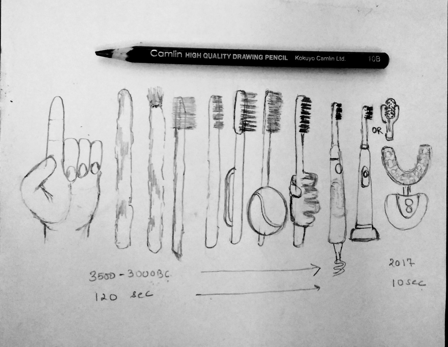
AI Augmented Brushing For Special Needs

Disorder of neural development contributes to the lack of necessary manual dexterity. These lack of fine motor skills adds up to the difficulties of care givers and accounts for poor oral health among children with special needs.

Regular manual tooth brush depend on the user’s controlled movements but the presence of physical and cognitive disabilities among special needs can create difficulties in both holding as well as manipulating a tooth brush. Powered toothbrush on the other hand provide controlled pressure and movement along with time assisted technology.

These electronic tooth brush provide vibration and oscillation or mixed movement that effectively removes plaque and inhibit certain harmful bacteria affecting gingival health. Sonic vibration have been studied to stimulate salivary flow in xerostomia (including those induced by medication).

Different head sizes are powered to accomplished cleaning from soft, medium to hard brushing among different age group. Hard bristles are mostly recommended to patient with special needs, as they allow shorter brushing time and less application of pressure during brushing. Brush head in the powered brushes are compatible with better grasping efficiency among children with special needs.

However brushing with powered tooth brush has been associated to trigger the external stimulus among children with reflex epilepsy and therefore there use is warranted in such patient.

An element of safety puts these powered toothbrush at par with the manual toothbrush as the bristles are attached securely to the head, unlike filaments and even parts of manual brush which are reported to cause gingival and intra oral injuries following trauma.

Researchers conclude that powered tooth brush can remove 60% more plaque. As suggested by Cochrane study where powered tooth brush were compared to manual tooth brush regarding plaque removal, health of gingiva, staining and calculus, only powered toothbrush with rotation -oscillation action provided advantage.

The Artificial Intelligence (AI) assisted technology is the next step to the future of brushing. This basically requires you to guide along the tooth surface with 3D Mouth Mapping in Real time. The sensor in the brushes analyses brushing in real time and monitor progress through a mobile application and even allows you to cover those underbrush surfaces commonly skipped routinely. The assisted device will not only provide with different brushing techniques, pressure combination on various tooth surface but will also detect the change in oral environment and plaque accumulation,notifying you via an alarm to take steps for a better oral hygiene.

Create your own website, free on WordPress.com.Create your own website.
Start free